
How a CT Scan Works
A CT scan creates intricate cross-sectional images as opposed to a standard X-ray, which yields a single flat image. These CT images are formed by combining multiple X-ray images taken from different angles. This way, a CT scan allows clinicians to view the lungs in several thin slices, revealing the size, shape, density, and position of the abnormality with much greater precision.
In lung cancer care, CT scans serve two distinct roles: as a diagnostic tool when symptoms or an abnormal X-ray are present, and as a screening tool (LDCT) used annually in high-risk individuals to detect cancer before symptoms develop.
Standard CT vs. Low-Dose CT
- Standard CT is ordered when symptoms are present, a chest X-ray has shown something suspicious, or to stage a confirmed cancer. It uses a higher radiation dose and typically involves a contrast dye injected intravenously to improve the visibility of blood vessels and help differentiate a tumor from surrounding tissue.
- Low-dose CT uses significantly less radiation, approximately the same as a mammogram, with no contrast injection needed. It is appropriate as an annual screening test in high-risk individuals with no current symptoms and takes only a few minutes.
Detecting and Evaluating Lung Nodules
A central feature of CT screening is the detection and evaluation of lung nodules, small, rounded areas of tissue within the lung. According to a review in the Journal of Thoracic Imaging (Rubin, PMC4654704), detection of medium to large nodules on CT is consistent, but sensitivity decreases substantially as nodule size falls below 8 to 10 millimeters.
Most nodules found on CT are benign, often resulting from past infection or scarring. However, some require monitoring or further investigation to exclude malignancy.
What the Radiologist Looks For
When a CT scan reveals a nodule, the radiologist considers various factors to determine the possibility of cancer:
- Size: Larger nodules have a higher risk of being malignant. Those measuring less than 6 millimeters are generally considered very low risk.
- Shape and margins: Irregular or spiculated (spiky) margins indicate a greater risk as compared to smooth, well-defined borders.
- Density: Nodules that are solid, part-solid, or ground-glass. Early adenocarcinoma can look like part-solid or ground-glass nodules.
- Growth rate: A nodule that enlarges during follow-up imaging is more alarming than one that remains steady for two or more years.
- Location: Nodules in the upper lobes have a higher chance of being malignant.
These features are assessed using the Lung-RADS system (Lung CT Screening Reporting and Data System), which categorizes findings from Category 1 (negative or benign) to Category 4 (suspicious or tissue sampling required).
What Happens After a Suspicious CT Scan
A CT scan, no matter how suspicious, cannot confirm a lung cancer diagnosis. A tissue biopsy is always necessary to confirm the diagnosis. After a suggestive CT, the following step is usually a CT-guided needle biopsy, bronchoscopy, or endobronchial ultrasound (EBUS), depending on where the lesion is located. A PET-CT can also be used to determine whether a nodule has metabolic activity or if malignancy has spread.
What to Expect During a CT Scan
- Before the scan: Remove metal objects and wear a hospital gown. In case of contrast dye usage, avoid eating for a few hours beforehand.
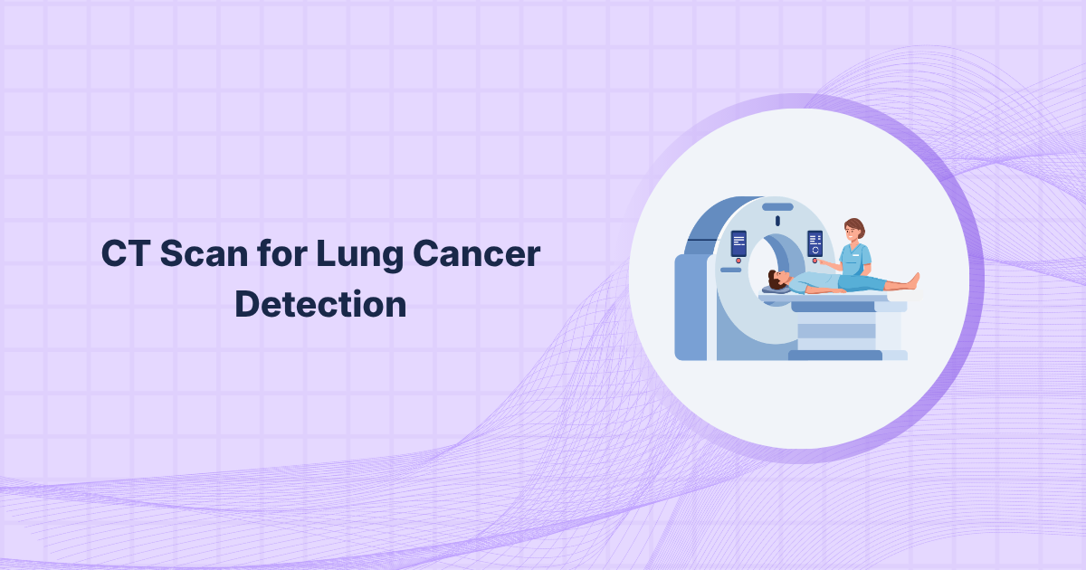
- During the scan: Lie on a table that slides into a ring-shaped machine. A low-dose CT takes under 10 minutes.
- Breath-hold instructions:You will be asked to briefly hold your breath to lessen the motion blur in the images.
- If contrast is used: You may feel a brief warm sensation or metallic taste. This clears naturally within hours.
- After the scan: Eat and drink normally. Results are typically reported to your doctor within a few days.
Conclusion
CT scans are very important for early diagnosis of lung abnormalities that could otherwise go undetected, particularly in individuals who are more prone to developing lung cancer. CT scans assist clinicians in identifying nodules early and determining if further monitoring or more targeted testing is required by offering comprehensive images of the lungs. Although a CT scan cannot diagnose cancer on its own, it is often the first important step in the diagnostic process. When used properly, it can greatly increase the likelihood of finding lung cancer at an earlier, more manageable stage.